Eversion of Foot Definition
What is eversion? An eversion definition: the movement in which the foot rotates so the sole faces away from the midline of the body. A simpler eversion meaning: if you were standing, eversion of the foot would occur if you rolled onto the inside portion of your foot causing the outside portion of your foot to rise off the ground.
What is inversion vs eversion of foot? Inversion of the foot can be thought of as the opposite movement of eversion. Inversion of the foot refers to the movement in which the foot rotates so the sole faces inward to the midline of the body. If you were standing, inversion of the foot would occur if you rolled onto the outside portion of your foot causing the inside portion of your foot to rise off the ground. Both eversion and inversion occur in the frontal plane, which involves side-to-side movements.

AnatomyFoot & Ankle
The foot and ankle form a complex system which consists of 28 bones, 33 joints, 112 ligaments, controlled by 13 extrinsic and 21 intrinsic muscles.
The foot is subdivided into the rearfoot, midfoot, and forefoot.
It functions as a rigid structure for weight bearing and it can also function as a flexible structure to conform to uneven terrain. The foot and ankle provide various important functions which includes:
Supporting body weight.
Providing balance.
Shock absorption.
Transferring ground reaction forces.
Compensating for proximal malalignment.
Substituting hand function in individuals with upper extremity amputation/paralysis.

StructureLigaments of the ankle medial aspect Primal.pngLigaments of the ankle posterior aspect Primal.png
The ankle or tibiotalar joint constitutes the junction of the lower leg and foot. The osseous components of the ankle joint include the distal tibia, distal fibula, and talus.
The anatomic structures below the ankle joint comprise the foot, which includes:
Hindfoot: Hindfoot, the most posterior aspect of the foot, is composed of the talus and calcaneus, two of the seven tarsal bones. The talus and calcaneus articulation is referred to as the subtalar joint, which has three facets on each of the talus and calcaneus.
Midfoot: The midfoot is made up of five of the seven tarsal bones: navicular, cuboid, and medial, middle, and lateral cuneiforms. The junction between the hind and midfoot is termed the Chopart's joint, which includes the talonavicular and calcaneocuboid joints.
Forefoot: The forefoot is the most anterior aspect of the foot. It includes metatarsals, phalanges (toes), and sesamoid bones. There are a metatarsal and three phalanges for each digit apart from the great toe, which only has two phalanges. The articulation of the midfoot and forefoot forms the Lisfranc joint.
Talocrural JointAnkleAxisPost

The tip of the medial malleoli is anterior and superior to the lateral malleoli, which makes its axis oblique to both the sagittal and frontal planes. The axis of rotation is approximately 13°-18° laterally from the frontal plane and at angle of 8°-10° from the transverse plane. Motion in other planes is required (like horizontal and frontal plane) to achieve a complete motion for plantarflexion and dorsiflexion.The reported normal available range for dorsiflexion varies in the literature between 0°-16.5°[8] and 0°-25°,[9] and this changes with weightbearing. The normal range of plantarflexion has been reported to be around 0°- 50°.
Gait and the Foot8 phases of gait cycle.png
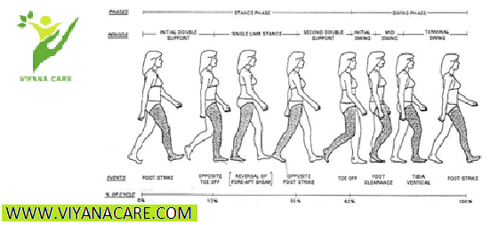
Gait is made up of repetitive cycles of the stance phase when the foot is on the ground (foot strike, mid stance, and terminal stance) and the swing phase when the foot is in the air. When running, there is an additional phase: the float phase when both feet are off the ground.
During walking, In foot strike, the foot is supinated, and Chopart joint is locked, making the foot rigid when the heel first lands.
The foot pronates and flattens during mid-stance as it comes in full contact with the surface.
Terminal stance is then characterized by propulsion via heel off and toe-off.
The Lisfranc joint allows slight dorsiflexion and plantarflexion.
Force is then transferred to the middle column of the forefoot during the toe-off phase of stepping, and the forefoot supinates.
The lateral column acts during the final phase of push-off while stepping, providing primarily sensory input.
The base of the fifth metatarsal alone absorbs significant force and weight.
The combination of fixed midfoot, slightly flexible Lisfranc joint, and flexible metatarsophalangeal joints create a lever for propulsion during gait[4].
Influence on Kinetic Chain/Gait
As discussed above with MT joint locking, the transition in the foot from pronation to supination is an important function that assists in adapting to uneven terrain and acting as a rigid lever during push off.
During pronation, the MT joint unlocks, providing flexibility of the foot and assisting in maintaining balance.
During supination, the MT joint locks, providing rigidity of the foot and maximizing stability.
If the foot remains pronated, it would lead to hypermobility of the midfoot and place greater demand on the neuromuscular structures that stabilize the foot and maintain upright stance. Whereas if the foot remains supinated, the midfoot would be hypomobile, which would compromise the ability of the foot to adjust to the terrain and increase demand on surrounding structures to maintain postural stability and balance. Cote et al.[17] concluded that postural stability is affected by foot position in both static and dynamic conditions. Chain reactions occur secondary to the positioning of the foot.
In closed chain movements, the following kinetic chain reaction takes place in an over-pronated foot:
Kinetic chain.png
Calcaneal eversion
Adduction and plantarflexion of talus
Medial rotation of talus
Medial rotation of tibia and fibula
Valgus at knee
Medial rotation of femur
Anterior tilting of pelvis
In closed chain movement the following kinetic chain reaction takes place in an over-supinated foot:

Calcaneal inversion
Abduction and dorsiflexion of talus
Lateral rotation of talus
Lateral rotation of tibia and fibula
Varus at knee
Lateral rotation of femur
Posterior tilting of pelvis
FOOT EVERSION SIGNS, CAUSES AND TREATMENT
OUR EXPERTS EXPLAIN THE FACTS ON FOOT EVERSION
Foot eversion is when your foot collapses inward, usually with your feet also flattening. The sole of the foot actually faces away from your other foot, increasingly so as the problem worsens. Foot eversion is not particularly common among most people, although our experts often see athletes suffering from this type of foot pain. Many people think foot eversion is normal; it is not. However, it can be relatively easy to correct.
FOOT EVERSION CAUSES
Our experts, WI most commonly find foot eversion in those who stand in a pronated position (with the feet on an outward angle). This posture makes the body much weaker and more exposed. By itself, this might cause injury but it also results in less leverage and strength. In sports, this is a big limitation.
If foot eversion is not properly treated, it can cause other problems which are often far more painful.
Untreated foot eversion can cause plantar fasciitis (heel pain), ankle sprains, fractures or breaks, knee problems, bone spurs, Achilles tendonitis and IT band problems.
FOOT EVERSION TREATMENT
Foot eversion treatment options generally include custom orthotics, braces or shoe inserts; however, the most common foot eversion treatment is physical therapy. Physical therapy is important to improve both the flexibility and strength of your foot and ankle muscles. Stronger, “corrected” muscles prevent injury and correct misalignment issues.
If you suffer from foot eversion, it is important to see our team.
What is eversion? An eversion definition: the movement in which the foot rotates so the sole faces away from the midline of the body. A simpler eversion meaning: if you were standing, eversion of the foot would occur if you rolled onto the inside portion of your foot causing the outside portion of your foot to rise off the ground.
What is inversion vs eversion of foot? Inversion of the foot can be thought of as the opposite movement of eversion. Inversion of the foot refers to the movement in which the foot rotates so the sole faces inward to the midline of the body. If you were standing, inversion of the foot would occur if you rolled onto the outside portion of your foot causing the inside portion of your foot to rise off the ground. Both eversion and inversion occur in the frontal plane, which involves side-to-side movements.
AnatomyFoot & Ankle
The foot and ankle form a complex system which consists of 28 bones, 33 joints, 112 ligaments, controlled by 13 extrinsic and 21 intrinsic muscles.
The foot is subdivided into the rearfoot, midfoot, and forefoot.
It functions as a rigid structure for weight bearing and it can also function as a flexible structure to conform to uneven terrain. The foot and ankle provide various important functions which includes:
Supporting body weight.
Providing balance.
Shock absorption.
Transferring ground reaction forces.
Compensating for proximal malalignment.
Substituting hand function in individuals with upper extremity amputation/paralysis.
StructureLigaments of the ankle medial aspect Primal.pngLigaments of the ankle posterior aspect Primal.png
The ankle or tibiotalar joint constitutes the junction of the lower leg and foot. The osseous components of the ankle joint include the distal tibia, distal fibula, and talus.
The anatomic structures below the ankle joint comprise the foot, which includes:
Hindfoot: Hindfoot, the most posterior aspect of the foot, is composed of the talus and calcaneus, two of the seven tarsal bones. The talus and calcaneus articulation is referred to as the subtalar joint, which has three facets on each of the talus and calcaneus.
Midfoot: The midfoot is made up of five of the seven tarsal bones: navicular, cuboid, and medial, middle, and lateral cuneiforms. The junction between the hind and midfoot is termed the Chopart's joint, which includes the talonavicular and calcaneocuboid joints.
Forefoot: The forefoot is the most anterior aspect of the foot. It includes metatarsals, phalanges (toes), and sesamoid bones. There are a metatarsal and three phalanges for each digit apart from the great toe, which only has two phalanges. The articulation of the midfoot and forefoot forms the Lisfranc joint.
Talocrural JointAnkleAxisPost
The tip of the medial malleoli is anterior and superior to the lateral malleoli, which makes its axis oblique to both the sagittal and frontal planes. The axis of rotation is approximately 13°-18° laterally from the frontal plane and at angle of 8°-10° from the transverse plane. Motion in other planes is required (like horizontal and frontal plane) to achieve a complete motion for plantarflexion and dorsiflexion.The reported normal available range for dorsiflexion varies in the literature between 0°-16.5°[8] and 0°-25°,[9] and this changes with weightbearing. The normal range of plantarflexion has been reported to be around 0°- 50°.
Gait and the Foot8 phases of gait cycle.png
Gait is made up of repetitive cycles of the stance phase when the foot is on the ground (foot strike, mid stance, and terminal stance) and the swing phase when the foot is in the air. When running, there is an additional phase: the float phase when both feet are off the ground.
During walking, In foot strike, the foot is supinated, and Chopart joint is locked, making the foot rigid when the heel first lands.
The foot pronates and flattens during mid-stance as it comes in full contact with the surface.
Terminal stance is then characterized by propulsion via heel off and toe-off.
The Lisfranc joint allows slight dorsiflexion and plantarflexion.
Force is then transferred to the middle column of the forefoot during the toe-off phase of stepping, and the forefoot supinates.
The lateral column acts during the final phase of push-off while stepping, providing primarily sensory input.
The base of the fifth metatarsal alone absorbs significant force and weight.
The combination of fixed midfoot, slightly flexible Lisfranc joint, and flexible metatarsophalangeal joints create a lever for propulsion during gait[4].
Influence on Kinetic Chain/Gait
As discussed above with MT joint locking, the transition in the foot from pronation to supination is an important function that assists in adapting to uneven terrain and acting as a rigid lever during push off.
During pronation, the MT joint unlocks, providing flexibility of the foot and assisting in maintaining balance.
During supination, the MT joint locks, providing rigidity of the foot and maximizing stability.
If the foot remains pronated, it would lead to hypermobility of the midfoot and place greater demand on the neuromuscular structures that stabilize the foot and maintain upright stance. Whereas if the foot remains supinated, the midfoot would be hypomobile, which would compromise the ability of the foot to adjust to the terrain and increase demand on surrounding structures to maintain postural stability and balance. Cote et al.[17] concluded that postural stability is affected by foot position in both static and dynamic conditions. Chain reactions occur secondary to the positioning of the foot.
In closed chain movements, the following kinetic chain reaction takes place in an over-pronated foot:
Kinetic chain.png
Calcaneal eversion
Adduction and plantarflexion of talus
Medial rotation of talus
Medial rotation of tibia and fibula
Valgus at knee
Medial rotation of femur
Anterior tilting of pelvis
In closed chain movement the following kinetic chain reaction takes place in an over-supinated foot:
Calcaneal inversion
Abduction and dorsiflexion of talus
Lateral rotation of talus
Lateral rotation of tibia and fibula
Varus at knee
Lateral rotation of femur
Posterior tilting of pelvis
FOOT EVERSION SIGNS, CAUSES AND TREATMENT
OUR EXPERTS EXPLAIN THE FACTS ON FOOT EVERSION
Foot eversion is when your foot collapses inward, usually with your feet also flattening. The sole of the foot actually faces away from your other foot, increasingly so as the problem worsens. Foot eversion is not particularly common among most people, although our experts often see athletes suffering from this type of foot pain. Many people think foot eversion is normal; it is not. However, it can be relatively easy to correct.
FOOT EVERSION CAUSES
Our experts, WI most commonly find foot eversion in those who stand in a pronated position (with the feet on an outward angle). This posture makes the body much weaker and more exposed. By itself, this might cause injury but it also results in less leverage and strength. In sports, this is a big limitation.
If foot eversion is not properly treated, it can cause other problems which are often far more painful.
Untreated foot eversion can cause plantar fasciitis (heel pain), ankle sprains, fractures or breaks, knee problems, bone spurs, Achilles tendonitis and IT band problems.
FOOT EVERSION TREATMENT
Foot eversion treatment options generally include custom orthotics, braces or shoe inserts; however, the most common foot eversion treatment is physical therapy. Physical therapy is important to improve both the flexibility and strength of your foot and ankle muscles. Stronger, “corrected” muscles prevent injury and correct misalignment issues.
If you suffer from foot eversion, it is important to see our team.